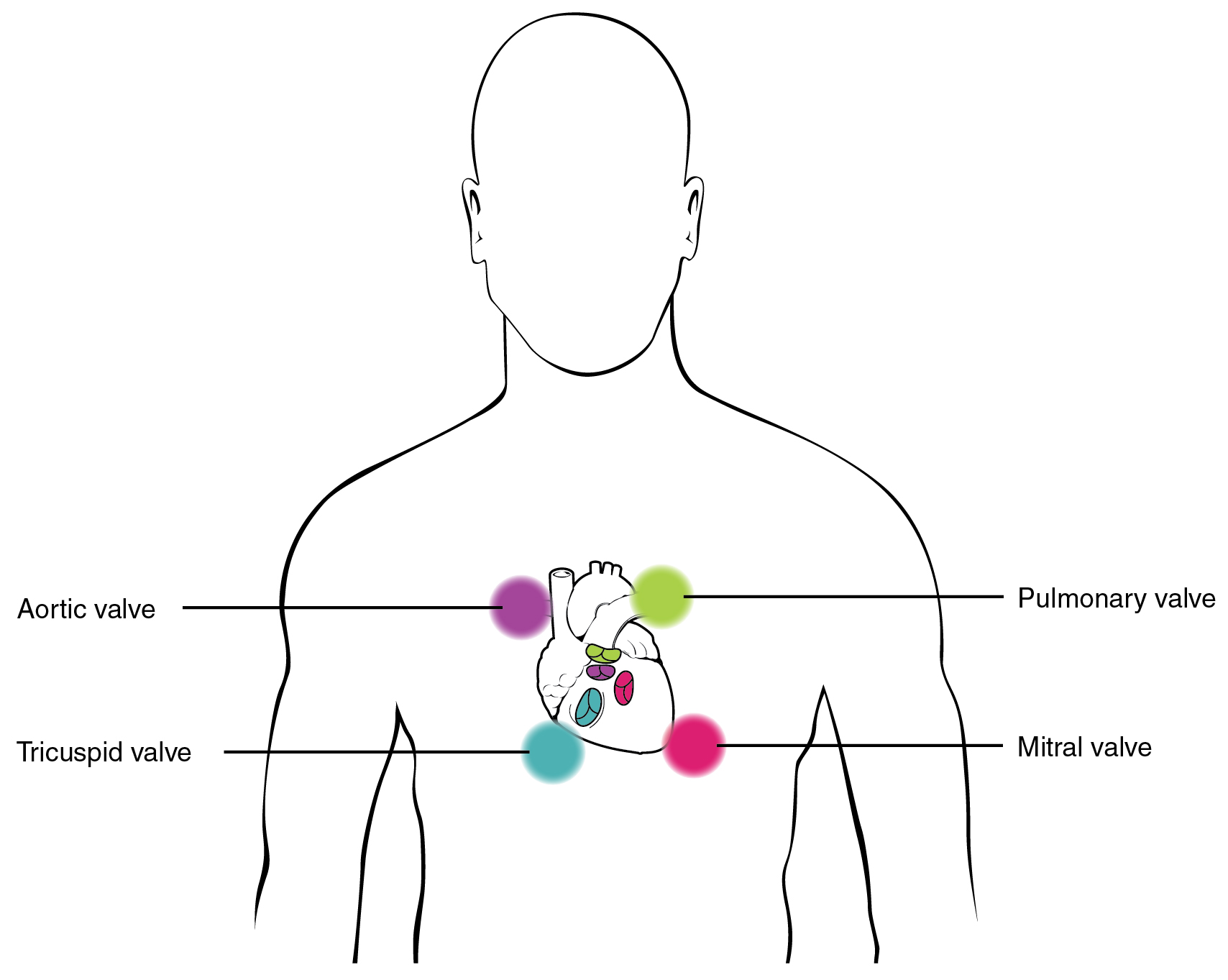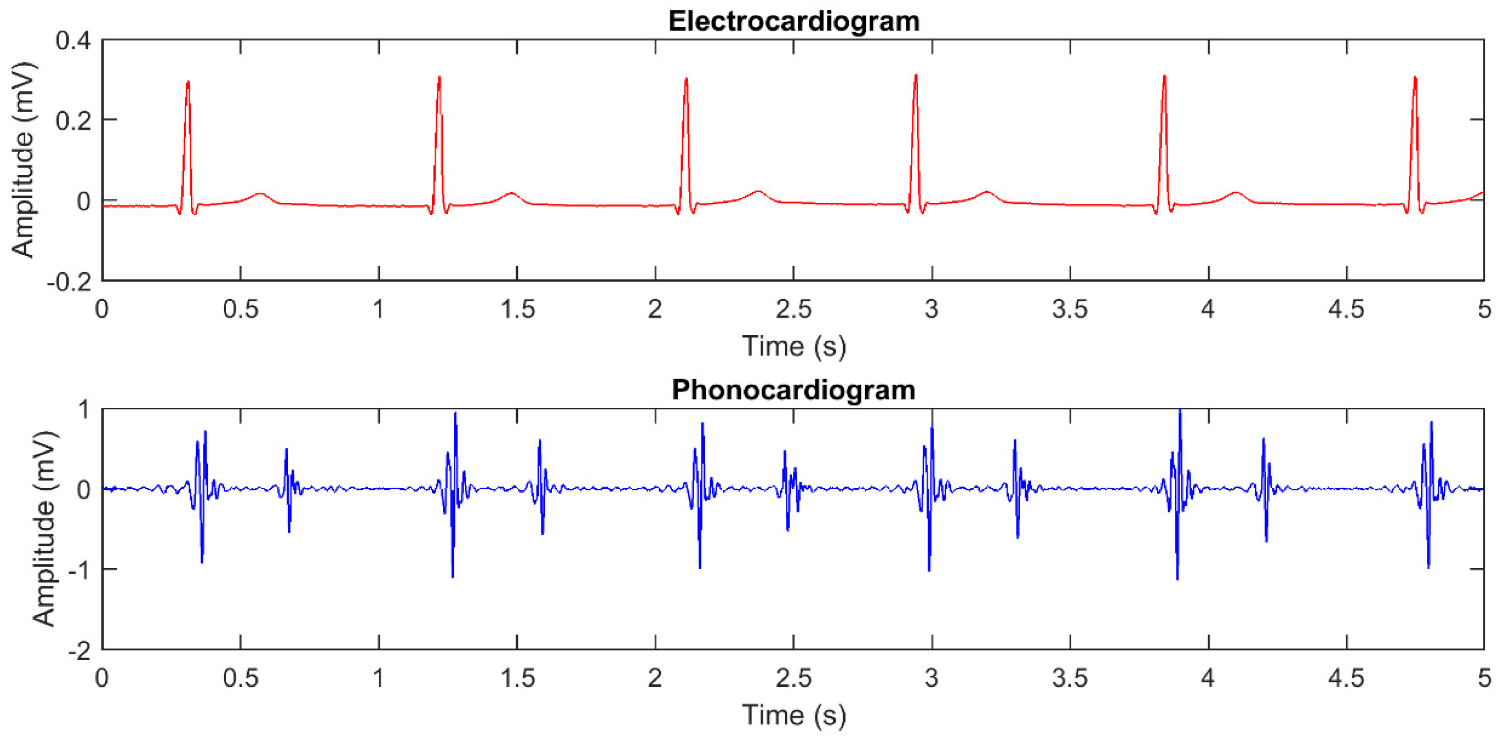

We use “ba” as the first part of the word to indicate that the first beat is always shorter in length than the second. “Ba boom” is a great way to use onomatopoeia a bit more specifically than “thump thump.” We can use two varying sounds instead to show that there is a specific way for a heart to make a beating sound. Thump thump! Thump thump! Can you hear my heartbeat? I feel like it’s being really loud right now! Ba Boom.Thump thump! Thump thump! I never thought she was going to ask me out like that!.Thump thump! Thump thump! My heart is racing right now.It’s also a very popular choice for many people to help them give a more tactile idea of what the sound is (since they can “thump” something to demonstrate the sound). “Thump thump” is great because it shows that there’s a drumming beat with the heart. Expecting parents can conveniently listen to their baby’s heartbeat in the comfort of their home.Watch the video: Only 1 percent of our visitors get these 3 grammar questions right. Nowadays, it is widely accessible since it is possible to buy a Doppler machine at a modest cost for personal use. The Doppler used to be reserved for health professionals. The Doppler can be used on its own, but it is often included in the sonography machine. The instrument emits ultrasounds to detect blood flow in the heart vessels. The obstetrician adds the exam to prenatal visits starting on the 10 th or 11 th week of pregnancy, to check on the baby’s strength and health. While the sonogram allows us to see the baby’s heart, the Doppler allows us to hear it. During his initial experimentation, the procedure took place in a bathtub to prevent the sound waves from escaping into thin air. The first obstetric sonogram was performed by the British Ian Donald in 1958. During the 1950’s, the technology entered the medical field with sonography equipment that could probe inside the human body. They helped detect submarines during World War One, or locate icebergs. Ultrasounds were first used in the sailing world at the beginning of the 20 th century. On the sonogram image, the heartbeat is indicated by a flickering spot. These sound waves reflect on the fetus, allowing us to study its organs, keep an eye on its growth, discover potential anomalies and determine its sex. Sonography: a Window into the Human Bodyīased on the same principle as the sonar, the obstetric sonogram works by sending ultrasounds into the uterus.


The sound could only be perceived beyond the 18 th week of pregnancy. Similar to Laennec’s stethoscope, which consisted of a wooden cone with a metal plate at one end, the instrument was applied to the mother’s stomach to listen to her child’s heartbeat. Before that, starting at the beginning of the 19 th century, the obstetric stethoscope was used to hear the baby’s heart. This technique became widespread in the 1970’s. Nowadays, it is possible to detect the fetal heartbeat as early as the fifth or sixth week of pregnancy thanks to the sonogram. During the following weeks, the heart keeps growing and developing to form its four ventricles. At this stage, it is already pumping blood throughout the baby’s body. The embryo is then barely two to three millimetre long, the size of an apple seed. The human heart starts to beat as early as the 21 st day of pregnancy.


 0 kommentar(er)
